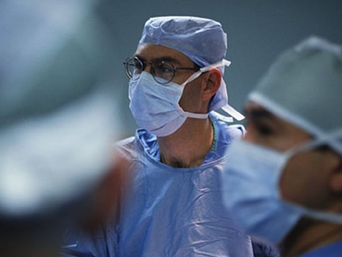
MONDAY, Jan. 6, 2020 (HealthDay News) -- A novel workflow that combines advanced optical imaging with an artificial intelligence algorithm may accurately diagnose brain tumors in real time in the operating room, according to a study published online Jan. 6 in Nature Medicine.
Todd C. Hollon, M.D., from University of Michigan in Ann Arbor, and colleagues developed a parallel workflow that combines stimulated Raman histology (SRH), a label-free optical imaging method, and deep convolutional neural networks (CNNs) to predict diagnosis at the bedside in near real time in an automated fashion. The authors prospectively tested the workflow in a clinical trial of 278 patients with brain tumors.
The researchers found that CNN-based diagnosis of SRH images was noninferior to pathologist-based interpretation of conventional histologic images (overall accuracy, 94.6 versus 93.9 percent). The workflow predicted brain tumor diagnosis in the operating room in <150 seconds, an order of magnitude faster than conventional techniques, which take an estimated to 20 to 30 minutes.
"As surgeons, we're limited to acting on what we can see; this technology allows us to see what would otherwise be invisible, to improve speed and accuracy in the operating room, and reduce the risk of misdiagnosis," a coauthor said in a statement. "With this imaging technology, cancer operations are safer and more effective than ever before."
Several authors disclosed financial ties to Invenio Imaging, a company developing SRH microscopy systems.
Abstract/Full Text (subscription or payment may be required)