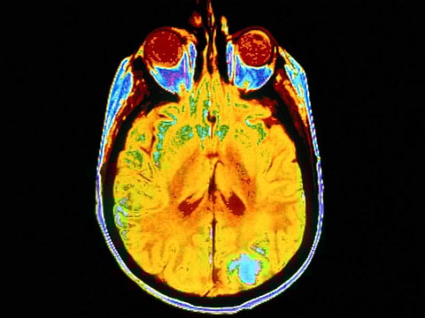
THURSDAY, May 6, 2021 (HealthDay News) -- A deep learning model is feasible for differentiating normal or likely abnormal brain magnetic resonance imaging (MRI), according to a study published online April 21 in Radiology: Artificial Intelligence.
Romane Gauriau, Ph.D., from the MGH & BWH Center for Clinical Data Science in Boston, and colleagues conducted a retrospective study to develop a deep learning approach using T2-weighted fluid attenuated inversion recovery (FLAIR) images to classify brain MRI as likely normal or likely abnormal. A convolutional neural network was trained on a large heterogeneous dataset covering a broad range of pathologies. Data were included from three datasets: Dataset A included 2,839 patients; dataset B included 6,442 patients; and dataset C included 1,489 patients. Datasets A and B were split into training, validation, and test sets, while dataset C was only used for testing.
The researchers found that the F1-score was 0.72 for model A, which was trained on dataset A from one institution and tested on dataset C from another institution; when compared with findings from the radiologic reports, the area under the curve was 0.78.
"The problem we are trying to tackle is very, very complex because there are a huge variety of abnormalities on MRI," Gauriau said in a statement. "We showed that this model is promising enough to start evaluating if it can be used in a clinical environment."
One author disclosed financial ties to the medical device industry.
Abstract/Full Text (subscription or payment may be required)