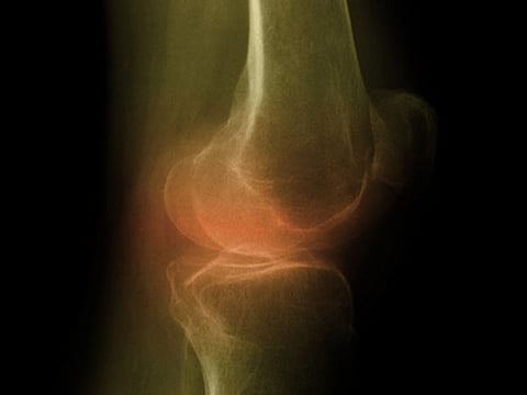
TUESDAY, Aug. 8, 2017 (HealthDay News) -- Bone mineral density (BMD), particularly in the hip, is negatively associated with knee cartilage defects and bone marrow lesions (BMLs) in patients with knee osteoarthritis, according to a study published online Aug. 1 in the International Journal of Rheumatic Diseases.
Qicui Zhu, from the First Affiliated Hospital of Anhui Medical University in Hefei, China, and colleagues conducted T2-weighted fast spin echo magnetic resonance imaging to assess knee cartilage defects and BMLs in 185 participants with symptomatic knee osteoarthritis. Dual-energy X-ray absorptiometry measured total body, hip and spine BMD.
The researchers found that after adjusting for potential confounders, total hip BMD was negatively associated with medial tibial cartilage defects, lateral femoral cartilage defects, medial tibial BMLs, and lateral tibial BMLs. There was a negative association between spine and total body BMD with lateral femoral cartilage defects, but not with BMLs.
"We concluded that BMD, particularly at the hip, was negatively associated with knee cartilage defects and BMLs," the authors write.
Abstract
Full Text (subscription or payment may be required)